The anatomy of the human heart: structure and function
Where is the human heart located in the body? And how does it work? Find out how the organ is structured, what tasks it performs and what roles its individual components play.
Where is the heart located?
Clench your hand into a fist. Your heart is about that size. Move your hand to your breastbone and then slightly to the left. Your heart sits there, in your ribcage, slightly offset between your two lungs. Its shape resembles a cone that tapers downwards and adjoins the diaphragm. This powerful muscle pump drives blood throughout the body and supplies organs and tissues.
What does the anatomy of the heart look like?
The heart is a hollow muscle and weighs around 300 grams. Its anatomy is shaped by muscle tissue and solid connective tissue. Discover the individual components and their functions in more detail.
Sides, atria and ventricles
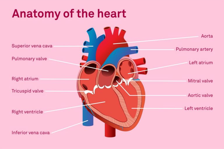
A muscular septum divides the left side of the heart from the right, separating oxygen-rich and oxygen-poor blood. Each half has two chambers – an atrium at the top and a ventricle at the bottom. This creates a total of four chambers, namely the right and left atria and the right and left ventricles.
The atrium and ventricle perform different functions in the heart. The two atria collect the incoming blood and send it downwards, before the ventricles actually pump it around.
Heart wall and pericardium
All four chambers are surrounded by the heart wall. This consists of three layers:
- Endocardium: the inner layer lines the heart chambers.
- Myocardium: the middle layer consists of cardiac muscles.
- Epicardium: the outer layer envelops the surface of the heart.
The pericardium covers the heart wall. It consists of connective tissue and surrounds the entire heart. There is a narrow fluid-filled space between the epicardium and the pericardium. With each heartbeat, the heart contracts and expands. The fluid allows it to glide gently in the pericardium and protects it from friction.
Cardiac skeleton and heart valves
Inside the heart, a structure formed of solid connective tissue – the cardiac skeleton – provides support. Located between the atria and the ventricles, it anchors the four heart valves with stable fibrous rings.
The four heart valves only let the blood flow in one direction. They are divided into two types. The two atrioventricular valves are located between the atria and the ventricles. On the left is the mitral valve, on the right the tricuspid valve. The two semilunar valves are located at the exit of the ventricles to the large arteries and are known as the aortic valve and the pulmonary valve.
Coronary arteries
The heart muscle also needs oxygen and nutrients, which is why the heart has its own blood vessels. They extend like a crown over the outside of the heart and are therefore called coronary arteries. Starting from the aorta, they divide into a left and a right coronary artery. Both branch out finer and finer to supply the entire heart. If a coronary artery becomes narrowed or blocked, not enough blood reaches the heart muscle. This can lead to a heart attack.
How does the heart work?
Together, the blood vessels and the heart form the cardiovascular system. The heart pumps blood through two circuits: the small lung circuit and the large body circuit.
When the heart muscle relaxes, oxygen-rich blood flows from the left atrium into the left ventricle. At the same time, oxygen-poor blood flows from the right atrium into the right ventricle. When the muscle tenses again, the left ventricle pumps blood into the aorta and the right ventricle pumps blood into the pulmonary artery. From the aorta, the blood flows to the body’s cells via arteries, arterioles and capillaries. Oxygen and nutrients are then released to the tissues. At the same time, the blood absorbs carbon dioxide and “waste materials”. The oxygen-poor blood then enters the right atrium via the body’s veins. From there, it flows into the right ventricle.
What causes the heartbeat?
The heart triggers its own heartbeat with an electrical signal. This signal is generated in the sinus node, the natural pacemaker in the right atrium. The sinus node consists of specialised, modified heart cells and emits electrical impulses on a regular basis. Each impulse first stimulates the two atria to contract.
From there, the signal continues to the AV node (atrioventricular node) at the border between the atria and ventricles. The cardiac skeleton made of connective tissue does not conduct electricity and separates the electrical signals of both areas. Therefore, the pulse only reaches the ventricles via the AV node. Fine fibres then conduct the signal to the heart muscle in the ventricles and stimulate it to tense up. This regular interplay of signal and tension determines the function of the heart.
Systoles and diastoles: how the heart pumps
Each heartbeat consists of two phases. During systole, the heart muscle contracts and pushes the blood out of the ventricles. This is why it is also known as the contraction and ejection phase. During diastole, the muscle relaxes and the ventricles fill up. This phase is therefore called the relaxation and filling phase.
The four heart valves open and close in the rhythm of these two phases. In systole, the semilunar valves open up the pathway to the large arteries. In diastole, the atrioventricular valves between the atria and the ventricles open. In this manner, the heart muscle and valves coordinate with each beat.
The path of blood through the body
The heart keeps the blood constantly moving through the body. In the arteries, oxygen-rich blood flows from the heart to the organs, while used-up, carbon-dioxide-rich blood flows back to the heart in the veins. The pulmonary artery and pulmonary vein are the exceptions. The former carries the carbon dioxide-rich blood to the lungs, while the latter carries oxygen-rich blood back from there.
From the right ventricle, blood flows into the pulmonary artery and on to the lungs. There, the pulmonary artery branches out into the fine capillaries around the alveoli. The blood releases carbon dioxide and absorbs fresh oxygen. It then travels back to the left atrium of the heart via the pulmonary vein.
At rest, the heart beats around 60 to 100 times per minute. Under exertion or stress, the frequency increases to 180 beats per minute so it can adapt its power to the body’s needs. Further facts about the heart reveal its power:
- The heart beats around 100,000 times every single day.
- It pumps roughly 6000 litres of blood through the body during this period.
- It moves approximately 5 litres of blood per minute.
- In the aorta, the blood flows at about 1 metre per second.
- Your heart takes several billion beats in your lifetime.
Blood pressure and heart health
With each beat, the heart pumps blood into the body. This results in blood pressure: the force that the blood exerts on the walls of the arteries, capillaries and veins.
A blood pressure reading always contains two numbers. The higher value relates to the moment when the heart contracts and pumps blood into the arteries. The lower one corresponds to the pressure as the heart relaxes and fills with blood. Balanced blood pressure that protects the heart and circulation is important for your health. Persistently high blood pressure puts the body under strain.
Illnesses of the cardiovascular system
Illnesses of the heart and blood vessels are widespread. Common cardiovascular diseases include heart failure, constricted coronary vessels, cardiac arrhythmia and high blood pressure. In severe cases, they can lead to a heart attack or stroke. The risk can be minimised with sufficient exercise, a balanced diet and reduced stress.
Now you know the anatomy of the heart and its functions in the body, you might be wondering how you can keep this vital organ healthy in the long term. We have compiled even more tips for a strong heart for you. In-depth information on illnesses and prevention is available from the Swiss Heart Foundation. Take your heart health seriously and help support your long-term well-being.

Dr Robert C. Keller provided the editorial team with advice. He is the Managing Director of the Swiss Heart Foundation, based in Bern, and has many years of experience in the field of cardiovascular illnesses. He runs the research department at the foundation.


Newsletter
Find out more about current health issues every month and get all the information you need about our attractive offers from all Helsana Group companies * delivered by e-mail to read whenever it suits you. Our newsletter is free of charge and you can sign up here:
We did not receive your information. Please try again later.
* The Helsana Group comprises Helsana Insurance Company Ltd, Helsana Supplementary Insurances Ltd and Helsana Accidents Ltd.